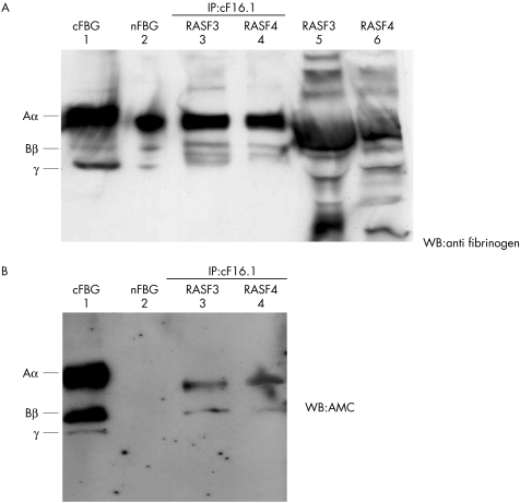
Figure 6 A demonstration of the presence of cFBG in RA synovial fluids using the immunoprecipitation (IP)‐western blotting (WB) method. Samples diluted 1:15 and immunoprecipitates using cF16.1 were obtained for cFBG positive RASFs (RASF3, RASF4) and subjected to western blotting by anti‐ fibrinogen (A) and AMC (B). Lane 1, cFBG as the positive control; lane 2, nFBG as the positive control for anti‐ fibrinogen and negative control for AMC; lanes 3 and 4, immunoprecipitates of RASF3 and 4 using cF16.1; lanes 5 and 6, RASF3 and 4 diluted at 1:15. Three consecutive chains at the levels of fibrinogen Aα, Bβ, γ were immunoprecipitated (A, lanes 3, 4) from many fibrinogen related products (A, lanes 5, 6), and Aα and Bβ chains in RASF 3, 4 were confirmed to be citrullinated. (B) The signal for AMC was stronger in Aα than in Bβ.

An official website of the United States government
Here's how you know
Official websites use .gov
A
.gov website belongs to an official
government organization in the United States.
Secure .gov websites use HTTPS
A lock (
) or https:// means you've safely
connected to the .gov website. Share sensitive
information only on official, secure websites.
