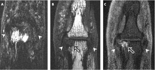
Figure 1 Entheseal‐related bone changes shown by (A) an osteoarthritic distal interphalangeal (DIP) joint scanned using T2‐weighted fat‐suppressed coronal sequence (B) a T1‐weighted coronal image of a different DIP joint and (C) the corresponding post‐gadolinium image showing a bone erosion that enhances after contrast (open arrows). (A) Thickened ligaments, which were degenerative bilaterally with fraying at the enthesis, can be seen (arrowheads). Prominent bone oedema is seen on the proximal phalange (arrow), which has high signal. This oedema has a relationship with the ligament that articulates with the adjacent bone as shown in fig 2. (B, C) As in the case of bone oedema, erosions are commonly related to the collateral ligaments being present adjacent to the ligament (arrow heads). This shows how the position of the collateral ligaments influences the expression of periarticular bone oedema and bone erosion in hand osteoarthritis.

An official website of the United States government
Here's how you know
Official websites use .gov
A
.gov website belongs to an official
government organization in the United States.
Secure .gov websites use HTTPS
A lock (
) or https:// means you've safely
connected to the .gov website. Share sensitive
information only on official, secure websites.
