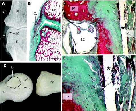
Figure 2 Radiological, osteological and histological images of the sites of attachment of the collateral ligaments of the distal interphalangeal (DIP) joints, showing the enthesis organ concept in relation to small‐joint osteoarthritis and how ligaments could effect bone oedema and erosion at these sites. (A) An anteroposterior radiograph of a joint from an elderly dissecting‐room cadaver printed to visualise the soft‐tissue relief of a collateral ligament (CL). The relatively proximal site of attachment of the ligament to the intermediate phalange (IP) means that the ligament presses against the lateral aspect of the bone in the region indicated by the arrow. BC, bone cyst; DP, distal phalange. (B) A low‐power view of a coronal histological section of a joint to show the area illustrated radiologically in (A). The thick collateral ligament presses against articular cartilage on the side of the intermediate phalange in the region of the arrow. (C) A collateral ligament leaves a smooth, well‐circumscribed marking (dotted line), devoid of vascular foraminae, on the intermediate phalange, which indicates the site of its enthesis (E). Consequently, in crossing the joint, the ligament presses against the side of the phalange in the region of the arrows. This area of bone is covered by a periosteal fibrocartilage that is continuous with the articular cartilage itself and is shown histologically in (B, C). The presence of fibrocartilage indicates that the bone is considerably compressed during ligament function at these sites. (D) A section in the coronal plane, through a collateral ligament of the proximal interphalangeal (PIP) joint in an elderly dissecting‐room cadaver. The head of the proximal phalange (PP) is covered with a periosteal fibrocartilage (PF) that articulates with the CL—the two are separated by the joint cavity (*) that extends around the side of the bone. The anatomical location of the bone cyst (BC) is underneath the periosteal fibrocartilage. Scale bar, 100 µm. (E) A higher power view of (D) to show the evidence of osteoarthritic degenerative change, fissuring and cell clustering (arrows), both in the periosteal fibrocartilage and in the collateral ligament. This supports the concept that the magnetic resonance imaging ligamentous abnormalities are caused by degenerative changes and are a key part of cartilage‐related disease in osteoarthritis. Scale bar, 100 µm.

An official website of the United States government
Here's how you know
Official websites use .gov
A
.gov website belongs to an official
government organization in the United States.
Secure .gov websites use HTTPS
A lock (
) or https:// means you've safely
connected to the .gov website. Share sensitive
information only on official, secure websites.
