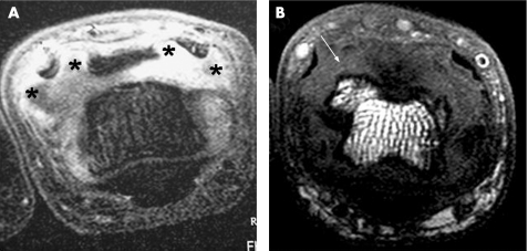
Figure 4 Formation of nodal osteoarthritis seen on axial magnetic resonance imaging scans of the proximal interphalangeal joints with early osteoarthritis in a patient with (A) symptom duration 5 weeks and (B) a more advanced early osteoarthritis joint. The joint in (A) scanned with gadolinium contrast was swollen with soft‐tissue swelling bulging dorsally, pushing the extensor tendon slips apart (*). Marked enhancement was seen, indicating active inflammation in the joint. An osteophyte was seen in the joint in (B) in the same area where the soft tissue tended to bulge through (arrow). This supports the hypothesis that the clinical features of nodal osteoarthritis, to some degree, mirror Baker's cyst formation in the knee joint.12

An official website of the United States government
Here's how you know
Official websites use .gov
A
.gov website belongs to an official
government organization in the United States.
Secure .gov websites use HTTPS
A lock (
) or https:// means you've safely
connected to the .gov website. Share sensitive
information only on official, secure websites.
