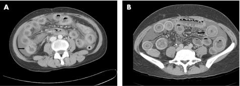
Figure 1 Computed tomography scans of patients with non‐recurrent (A) and recurrent (B) lupus enteritis. Both scans show ascites, mesenteric congestion and diffuse oedematous change in nearly whole halo viscous with engorged mesenteric vessels. The arrow indicates the thickest wall segment of the affected bowel.

An official website of the United States government
Here's how you know
Official websites use .gov
A
.gov website belongs to an official
government organization in the United States.
Secure .gov websites use HTTPS
A lock (
) or https:// means you've safely
connected to the .gov website. Share sensitive
information only on official, secure websites.
