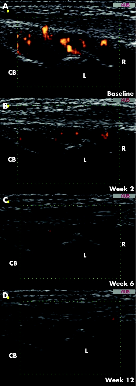
Figure 2 Representative power Doppler sonography (PDS) images obtained on longitudinal dorsal scan of the dominant wrist in a 62‐year‐old woman with a 2‐year history of disease (patient number 21). (A) Baseline PDS examination showed a marked increment in synovial tissue perfusion (PDS score 3). Follow‐up examinations at weeks (B) 2, (C) 6 and (D) 12 detected a clearly evident decrease in PDS signals (PDS score was 1, 0 and 0, respectively). CB, capitate bone; L, lunate bone; R, radius.

An official website of the United States government
Here's how you know
Official websites use .gov
A
.gov website belongs to an official
government organization in the United States.
Secure .gov websites use HTTPS
A lock (
) or https:// means you've safely
connected to the .gov website. Share sensitive
information only on official, secure websites.
