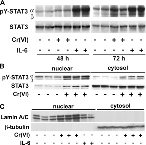
Figure 4. Subchronic exposure to Cr(VI) increases STAT3 phosphorylation.
(A) Total cell lysates were isolated from BEAS 2B cells exposed either to 5 μM Cr(VI) for 48 or 72 h or to 0.01 μg/ml of IL-6 for 30 min prior to the end of the experiment. Antibodies to pY-STAT3 and STAT3 were used in the Western blotting. (B) Cytosolic and nuclear protein fractions from BEAS 2B cells exposed for 72 h to 5 μM Cr(VI). Western-blot analysis was performed on 15 μg of nuclear protein or 30 μg of cytosolic protein. (C) The purity of the nuclear and cytosolic fractions was demonstrated by probing for the presence or absence of the nuclear protein, lamin A/C, or cytosolic β-tubulin. The experiments were performed in duplicate cell cultures and are representative of three independent experiments.

