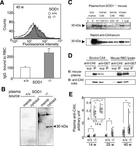
Figure 5. Production of autoantibodies to erythrocytes in SOD1-deficient mice.
(A) Detection of IgG bound to erythrocytes from SOD1−/− mice. Collected erythrocytes were reacted with FITC-labelled anti-mouse IgG followed by FACS analysis. Relative values of bound IgG are shown. n=7 for SOD1+/+ and n=5 for SOD1−/−. (B) Cytosolic and membrane fractions of RBCs were incubated with blood plasma from SOD1+/+ and SOD1−/− mice (1:50 dilution) followed by reaction with an HRP-conjugated anti-mouse IgG. (C) Immunoblot analysis using blood plasma from an SOD1−/− mouse and serum of a rabbit that had been immunized with bovine CAII. Western blot analysis of human CAII and RBCs with blood plasma from a SOD1−/− mouse at 40 weeks of age (1:50 dilution; upper panel). While mass marker b contains CAII, mass marker a does not. Characteristics of an anti-bovine CAII antibody (1:1000 dilution) raised in rabbit by immunoblotting of the same samples (lower panel); representative of several experiments. (D) Immunoprecipitation of bovine CAII and lysate of mouse RBCs with an anti-bovine CAII polyclonal antibody. Immunoblotting was performed with blood plasma from SOD1−/− mice and an anti-CAII monoclonal antibody (mAb). Representative data from several experiments. (E) The titres of the anti-CAII antibody in blood plasma were elevated, as judged by ELISA. The inset shows an enlargement of the results obtained for SOD1+/+ and SOD1−/− mice at 14 weeks of age.

