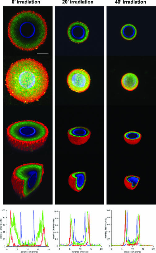
FIG. 7.
18B7 epitope distribution in the C. neoformans capsule. Cells were gamma irradiated for 0, 20, or 40 min and then labeled with 18B7-FITC. The cell wall was detected using calcofluor, and the capsular edge was detected by 12A1/goat anti-mouse IgM-TRITC. Pictures were taken using confocal microscopy. Panels show, for each period of gamma radiation, merged immunofluorescence labels, 3D reconstruction (ImageJ software), 3D z slice (Voxx software), 3D z/y slice (Voxx), and fluorescent signal intensity profiles (ImageJ) (top to bottom, respectively). Scale bar, 5 microns.

