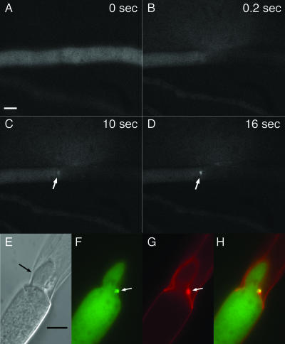
FIG. 3.
In AF-T8, SO-GFP accumulates sequentially at the septal plug and remains there after consolidation and reinitiation of tip growth. (A to D) Between 0 and 0.2 s, the hypha was cut with a UV pulse laser. After 10 s, SO-GFP accumulation becomes visible at the septal plug and intensifies during the next 6 s (white arrows) (see also movies S2 and S3 in the supplemental material). (E to H) Hyphae were cut with a razor blade and incubated for 30 min at room temperature prior to their analysis by light and fluorescence microscopy. A hypha from the adjoining compartment had formed a new tip, which grew into the dead compartment (black arrow). The new hypha is growing around the septal plug (white arrows). SO-GFP remains at the plug. (E) DIC image. (F) Fluorescent image with GFP filter. (G) Fluorescent image of calcofluor-stained hyphae. (H) Panels F and G merged. Bars represent 5 μm.

