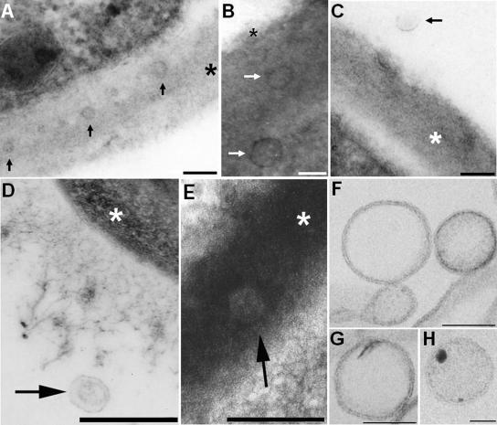
FIG. 1.
TEM of vesicles in acapsular (A to C) and encapsulated (D to H) C. neoformans cells. The occurrence of vesicles in association with the cell wall of acapsular cryptococci (A and B) or in the extracellular environment (C) is evident after in vitro growth. Vesicle-like structures were also observed in the lung following murine pulmonary infection (D and E). Putative vesicles near the edge of the capsule (D) or in the cryptococcal cell wall (E) were observed 2 h after infection. Bars, 100 nm (A to C) and 500 nm (D and E). Arrows point to vesicles, and asterisks are on the cryptococcal cell wall. (F to H) The pellets obtained by ultracentrifugation were isolated by differential centrifugation, purified from GXM by affinity chromatography, and analyzed by TEM. Extracellular vesicles with bilayered membranes and different profiles of electron density were observed. Bars, 100 nm (F and G) and 50 nm (H).

