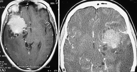
Fig. 1.

(A) MRI T1 weight (axial view) with gadolinium showing low density abnormalities (meningioma excised, patients L.F.) orbitofrontal cortex on the left side. (B) CT scan image showing low density abnormalities (meningioma excised, patients L.G.) orbitofrontal cortex on right sides
