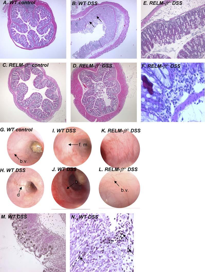
FIG 4.

Histopathology and colonoscopy in the colons of DSS-treated WT and RELM-β-/- mice. A-F, Representative photomicrographs of colons from control and DSS-treated WT and RELM-β-/- mice. G-L, Representative colonoscopy photographs of colons of control and DSS-treated WT and RELM-β-/- mice. M and N, Representative photomicrographs of immunohistochemically stained colon sections from DSS-treated WT mice using the RELM-β-specific polyclonal antiserum. Black arrows depict RELM-1 inflammatory cells. Black arrows in Fig 4, B, depict ulceration of the epithelial cell layer: black arrows in Fig 4, G and L, depict normal colonic vasculature (b.v., blood vessel); black arrows in Fig 4, I, H, and J, depict mucus and diarrhea (d), friable mucosa (f. m.), and rectal bleeding (b). Fig 4, H, Narrowing of colon, with evidence of a stricture. Original magnification: A-D, 350; E and M, 3100; F and N, 3200.
