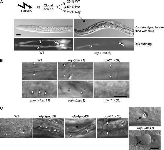
Figure 1.—
Mutagenesis and identification of rdy mutations. (A) Strategy for the identification of rdy mutations. After TMP/UV mutagenesis, the progeny of individual F1 animals were examined for the presence of rod-like larvae filled with fluid (top photos: DIC microscopy) and then for staining with the lipophilic dye DiO (bottom photos: fluorescence microscopy), which normally labels 12 neurons in the head (large arrows) and their dendrites (arrowheads). (Left) Wild-type (WT) larvae. (Right) rdy-1(mc38) mutants (the small arrow in the bottom photo points to the pharyngeal lumen). (B) L1 larva alae (arrowheads) of wild-type and rdy mutants viewed by DIC microscopy. Larvae are arranged in order of increased severity from WT = rdy-3 (normal alae) > rdy-2 > che-14 = rdy-4 > rdy-1 (no alae). (C) Excretory canal (open arrowheads) of L1 larvae viewed by DIC microscopy and arranged in order of increased severity: the canal was thicker in rdy-2 mutants; thicker and irregular (solid arrowhead) in rdy-4 mutants; extremely thick in rdy-1 mutants; essentially absent in rdy-3 mutants, in which vacuoles (arrow) that progressively grew in size (top—24 hr after egg laying; bottom—32 hr after egg laying) could be seen at the level of the excretory cell body. Bars, 5 μm.

