Figure 7.—
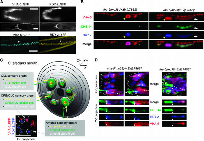
CHE-14, VHA-5, and RDY-2 co-accumulate in the epidermis of vha-5 mutants affecting secretion but not in their sensory organs. (A) Fluorescence micrographs showing VHA-5∷GFP (left) and RDY-2∷GFP (right) fusion proteins in different adults (top), or VHA-5∷CFP and RDY-2∷YFP fusion proteins in the same L2 larva (bottom). Both proteins are expressed in the same tissues, i.e., the amphid sheath cell (top), the excretory cell and canal (middle), the epidermis (bottom, arrowheads). (Top) Lateral view. (Bottom) Ventral views. Bars, 10 μm. (B) Confocal Z-projections through the epidermis (apical, top of each panel) showing the distributions of CHE-14∷YFP and RDY-2∷CFP in heterozygous (left) or homozygous (right) vha-5(mc38) adults carrying a vha-5(L786S)∷mrfp mutant transgene (denoted Ex[L786S]). All three fluorescent proteins accumulate and colocalize (white arrrow) in homozygous vha-5(mc38) animals; the che-14 transgene was generally too weak to be detected in the excretory canal (yellow arrowhead). (C) Scheme of the C. elegans mouth (for the sake of simplicity, IL sensory organs are not represented), showing sensory organs where VHA-5 and RDY-2 are expressed. The XZ projection of confocal micrographs (bottom left) shows partial colocalization of VHA-5 and RDY-2 in the apical sheath cells of the amphids (larger dotted circles) and CEP (small dotted circles) and/or in the sheath cells of the OLQ sensory organs (arrowheads); two of the four CEP/OLQ organs seem to have no red signal because it was more difficult to detect them on the other side of the animal. (D) XY projections of confocal micrographs through the head of heterozygous (left) or homozygous (right) vha-5(mc38) adults carrying a vha-5(L786S)∷mrfp mutant transgene, and YZ section through an amphid organ (corresponding to the dotted arrow in the XY projection). CHE-14 was uniquely expressed in the socket cell (to the left of the arrowhead; the sheath cell is to the right of the arrowhead). Five transgenic animals were examined in detail by confocal microscopy. Bars, 5 μm.

