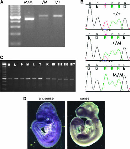
Figure 3.—
Identification of mutation in Tapt1 and RT–PCR analysis. (A) RT–PCR analysis of the Tapt1 gene using primers flanking exon 7. M, mutant (L5Jcs1 allele). Note the fainter mutant (lower) band in the heterozygote. The leftmost lane is a 100-bp ladder. (B) DNA sequence traces of indicated genotypes of the distal exon 7 splice junction. (C) PCR analysis of cDNAs from various tissues. (D) In situ hubridization of antisense or sense (control) RNAs to E9.5 wild-type embyros. H, heart; L, lung; B, brain; M, skeletal muscle; L, liver; T, testis; K, kidney; E, embryonic day.

