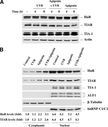
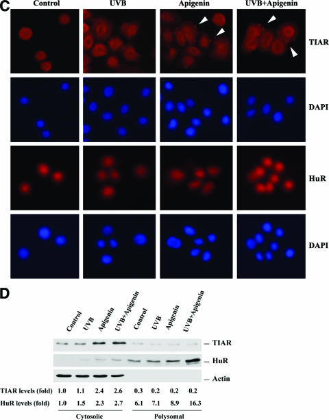
FIG. 5.
Effect of UVB and apigenin treatment on the subcellular location of HuR and TIAR. (A) Whole-cell lysates were prepared and subjected to Western blot analysis for HuR, TIAR, and TIA-1. Immediately after UVB (1,000 J/m2) or sham irradiation, apigenin was added to culture medium to a final concentration 50 μmol/liter, and cells were harvested at the indicated times. The “-” indicates untreated control. (B) Four hours after UVB or apigenin treatment, cytoplasmic and nuclear lysates were prepared and Western blot analysis was used to monitor the expression of HuR, TIAR, TIA-1, and AUF1. Expression levels of the cytoplasmic marker β-tubulin and the nuclear marker hnRNP C were also monitored. (C) The subcellular localizations of TIAR and HuR were also monitored by immunofluorescence, and stress granules (arrowheads) were observed in apigenin- and apigenin plus UVB-treated TIAR-stained cells. The slides were also stained with diamidinophenylindole for visualization of nuclei. (D)Western blot showing subcellular localization of TIAR and HuR in cytosol and polysomal fractions.


