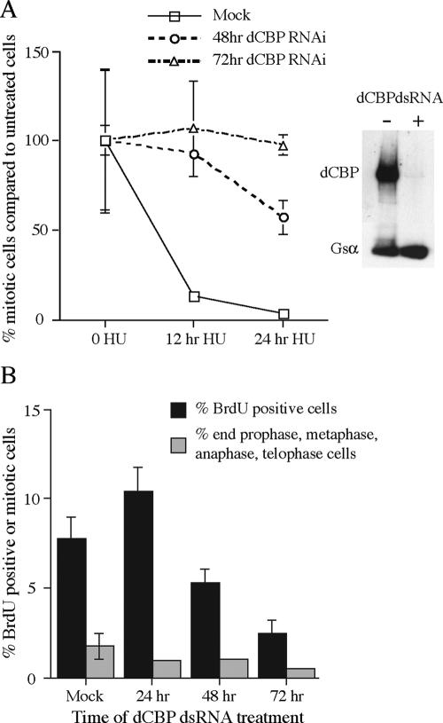
FIG. 4.
Loss of dCBP function causes a defect in the Kc cell replication checkpoint. (A) Kc cells treated with water (mock) or dCBP dsRNAs for 48 and 72 h were also treated with HU for 12 or 24 h before staining with the mitosis marker anti-phospho-H3 antibody. The mitotic index is expressed as a percentage of mitotic cells observed in the absence of HU treatment. Western analysis of mock-treated Kc cells or Kc cells treated with dCBP dsRNAs for 48 h shows that dCBP is depleted in the dsRNA-treated cells. Gsα was used as an internal control for gel loading and protein transfer. (B) Cells that are depleted of dCBP continue to incorporate BrdU, at least for 72 h. In addition, the frequency of mitotic cells (metaphase, anaphase, and telophase) was determined following phospho-H3 and propidium iodide staining.

