FIG. 1.
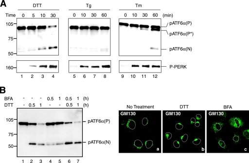
Effects of various ER stress inducers on activation of ATF6. (A) CHO cells were treated with 1 mM DTT, 600 nM Tg, or 4 μg/ml Tm for the indicated periods, and then cell lysates were prepared and analyzed by immunoblotting using anti-ATF6α or anti-phosphorylated PERK (P-PERK) antibodies. Migration positions of pATF6α(P), pATF6α(N), and P-PERK as well as full-range rainbow molecular weight markers (GE Healthcare) are indicated. pATF6α(P*) denotes the nonglycosylated form of pATF6α(P). (B) CHO cells were untreated (lane 1) or treated with 1 mM DTT alone (lanes 2 and 3), 5 μg/ml BFA alone (lanes 4 and 5), or 1 mM DTT and 5 μg/ml BFA simultaneously (lanes 6 and 7) for the indicated periods, and then cell lysates were prepared and analyzed by immunoblotting using anti-ATF6α antibody (left). CHO cells treated with 1 mM DTT or 5 μg/ml BFA for 1 h were analyzed by immunofluorescence using anti-GM130 antibody (right). An outline of the nucleus is indicated by a white line in each cell.

