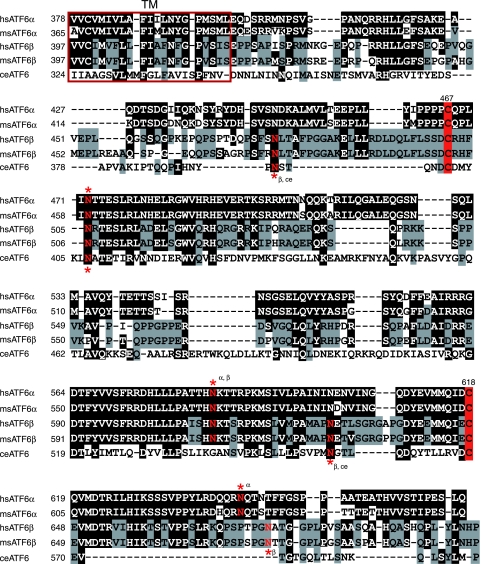
FIG. 2.
Alignment of the transmembrane and luminal domains of human and mouse ATF6α and ATF6β as well as worm ATF6. Amino acid sequences of the transmembrane (TM, red square) and luminal domains of human (hs) and mouse (ms) ATF6α and ATF6β as well as C. elegans (ce) ATF6 are aligned. Amino acids identical to those of ATF6α are marked by white letters in black boxes, whereas amino acids identical to those of ATF6β are shaded with gray. The two highly conserved cysteine residues are highlighted by red boxes. Asparagine residues marked by red and asterisks denote putative N-glycosylation sites.

