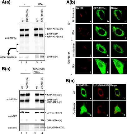
FIG. 8.
Effects of enforced expression of S1P in the ER on cleavage of ATF6. (A) (a) CHO cells transfected with pCMVshort-EGFP-ATF6α(WT) or pCMVshort-EGFP-ATF6α(C467&618A) were treated with or without 5 μg/ml BFA for 1 h. Cell lysates were prepared and analyzed under reducing conditions using anti-ATF6α antibody. The bottom panel shows the result of longer exposure of the region around pATF6α(N). (b) CHO cells transfected and treated as described for panel a were fixed and immunostained with anti-GM130 antibody (panels a, d, g, and j). GFP-ATF6α was visualized by its own fluorescence (panels b, e, h, and k). (B) (a) CHO cells were transfected with pCMVshort-EGFP-ATF6α(WT) or pCMVshort-EGFP-ATF6α(C467&618A) together with or without a plasmid to express S1P(ΔTMD)-KDEL tagged with the c-myc epitope at the C terminus. Cell lysates were prepared and analyzed under reducing conditions using anti-ATF6α (upper panel), anti-GFP (middle panel), or anti-myc (lower panel) antibodies. (b) CHO cells cotransfected as described for panel a were fixed and immunostained with anti-myc antibody (panels b and e). GFP-ATF6α was visualized by its own fluorescence (panels a and d).

