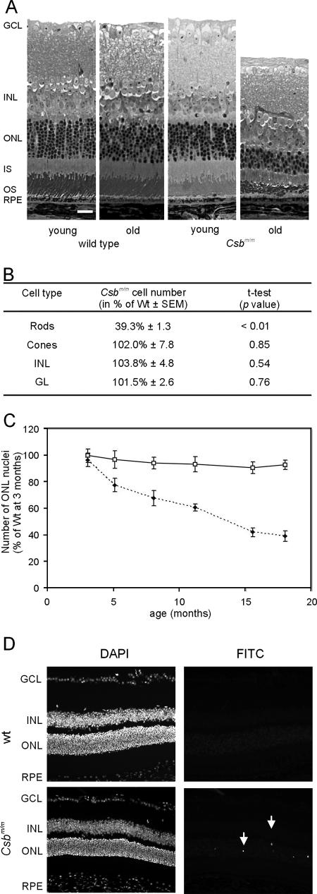
FIG. 2.
Progressive loss of photoreceptors with age in the Csbm/m mouse. (A) Representative micrographs of the central region of the retina of young (3 months) and old (18 months) wt and Csbm/m mice. Note the specific loss of ONL nuclei and distortion of the outer segment layer in the 18-month-old Csbm/m mouse. Bar, 25 μm. (B) Quantification of the number of nuclei in the various layers of the retina of 18-month-old wt and Csbm/m mice demonstrating a specific loss of rods in the aged Csbm/m retina (ANOVA, followed by t test). (C) Kinetics of photoreceptor cell loss in Csbm/m mice. The relative number of rod nuclei in wt and Csbm/m mice is plotted as the percentage of nuclei relative to that observed in 3-month-old wt mice. Open squares, wt; closed diamonds, Csbm/m. Error bars indicate standard deviations. (D) Micrographs of the retinas of 3-month-old wt and Csbm/m mice stained for apoptosis using the TUNEL method. Arrows indicate fluorescein isothiocyanate (FITC) staining for TUNEL-positive cells in the ONL (right panels). Nuclei were visualized using the DAPI (4′,6′-diamidino-2-phenylindole) staining method (left panels).

