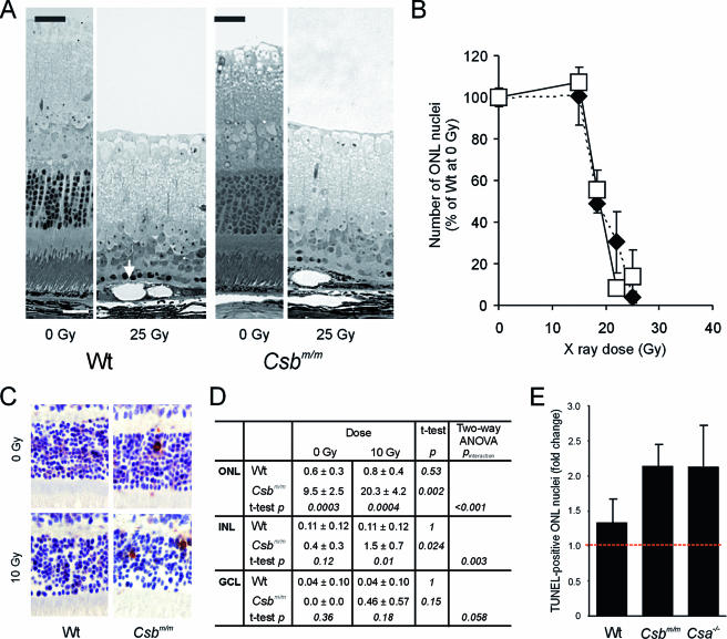
FIG. 4.
Hypersensitivity of the Csbm/m and Csa−/− retinas to ionizing radiation. (A) Micrographs of the retinas of wt and Csbm/m mice before and 7 days after focused exposure of the eye to a high dose of ionizing radiation (25 Gy; X rays). Note the nearly complete loss of photoreceptors in the exposed wt and Csbm/m retinas. The arrow indicates swollen RPE cells with large vacuoles. (B) Quantification of photoreceptor cell loss in wt and Csbm/m mice exposed to various doses (15 to 25 Gy) of X rays. ONL nuclei were counted in the peripheral retina and expressed as a percentage of the number of ONL nuclei present in the unexposed wt retina. Open squares, wt; closed diamonds, Csbm/m. (C and D) Effect of lower-dose (10 Gy; gamma rays) whole-body IR exposure to the retina of wt and Csbm/m mice (six animals/genotype, 8 to 10 weeks of age). (C) Example of apoptotic cells (recognized by a brown nucleus), as visualized by TUNEL staining. (D) Quantification of apoptotic (TUNEL-positive) cells in the retinal layers. Two-way ANOVA was used to test the independent effects of gamma rays and genotype on the number of TUNEL-positive cells as well as the interaction between these variables. (E) Comparison of ionizing radiation sensitivities of wt, Csbm/m, and Csa−/− retinas expressed as a change (n-fold) in the number of TUNEL-positive photoreceptor cells in the ONL of irradiated (10 Gy) versus unirradiated animals. Note the comparable hypersensitivities of Csa−/− and Csbm/m mice (two-way ANOVA; P value of interaction between genotype and irradiation is <0.001 for Csbm/m versus wt and 0.03 for Csa−/− versus wt).

