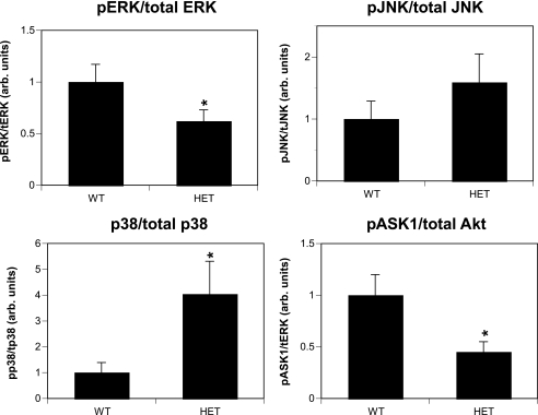
FIG. 4.
Analysis of signaling protein activity in 14-3-3τ+/− mice. Protein lysates were generated from ventricular tissue obtained from 10- to 12-week-old wild-type and 14-3-3τ+/− mice. Lysates were separated by SDS-PAGE and analyzed by immunoblotting with primary antibodies directed against phosphorylated p38, phosphorylated JNK, total JNK, phosphorylated ERK MAPK, total ERK MAPK, phosphorylated ASK1, phosphorylated Akt, and total Akt. Immunoblots were quantified by the computerized densitometry program Image J (NIH). Results were combined from three separate experiments (n = 11 for the wild type; n = 11 for 14-3-3τ+/− mice). arb. units, arbitrary units.

