FIG. 7.
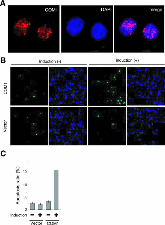
Restored COM1 protein localized in euchromatin and induced apoptosis in SYO-1 cells. (A) Localization of COM1 protein. Expression of COM1 in SYO-1 cells that conditionally express COM1 using the Tet-Off system. Clone #8 is shown at 72 h after doxycycline withdrawal. The signals were detected by indirect immunofluorescence with anti-FLAG antibody (COM1, left panel) and DAPI (middle panel). Merged signals (merge, right panel) showed that COM1 signals (red) were observed within the nucleus with a punctate pattern and in regions with little blue DAPI signal. (B) Apoptosis induction in SYO-1 cells by expression of COM1. Apoptosis was analyzed by caspase activity using a carboxyfluorescein FLICA apoptosis detection kit. The COM1-transfected SYO-1 cell (clone #8) is shown with doxycycline [induction(−)] and at 72 h after doxycycline withdrawal [induction (+)]. Empty vector-transfected SYO-1 cells (clone #4) are also shown as a control. The green signals are fluorescence of caspase-positive cells (left panel), and the blue signals are DNA visualized by Hoechst (right panel). (C) Average ratio of apoptosis (caspase-positive) cells depicted in Fig. 6B are shown. SYO-1 cells conditionally expressing COM1 showed a fourfold increase in apoptotic cell number 3 days after doxycycline withdrawal.

