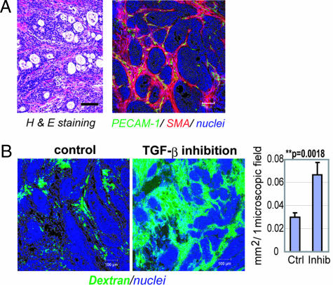
Fig. 1.
Histology of BxPC3 xenograft and effects of low-dose TβR-I inhibitor. (A) The histology of the TGF-β-nonresponsive BxPC3 xenograft, used as a model of poorly differentiated pancreatic adenocarcinoma, shown in H&E staining and immunohistochemistry. Examination revealed nests of tumor cells in gland-like structures, with areas rich in fibrotic components (filled by α-smooth muscle actin (SMA)-positive myofibroblasts, shown in red) between them. The tumor tissue also includes some PECAM-1-positive vessels (shown in green) in the interstitium, although almost no vasculature was observed inside the nests of tumor cells. (B) Dextran leakage. At 24 h after administration of low-dose TβR-I inhibitor (1 mg/kg i.p.), i.v.-administered dextran of 2 MDa (50 nm in hydrodynamic diameter) exhibited broader distribution with 1 mg/kg TβR-I inhibitor (Right) than in the control (Left), which was quantified and shown in the graph (n = 12). Error bars in the graphs represent standard errors, and P values were calculated by Student's t test. Ctrl, control; Inhib, inhibitor. (Scale bars, 100 μm.)

