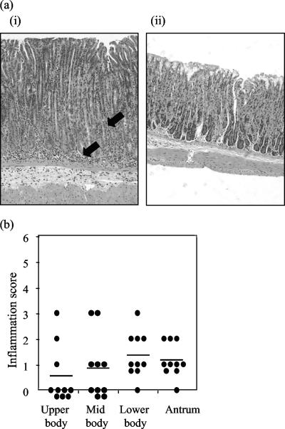
FIG. 2.
Histological examination of inflammation in the stomach. Stomach sections from mice infected for 6 months with H. pylori were stained with hematoxylin and eosin and analyzed for pathology (13). (a) Representative hematoxylin- and eosin-stained sections from infected (i) and uninfected (ii) mouse stomachs. Arrows indicate lymphocyte infiltration. (b) Summary of inflammation scores from various stomach regions of H. pylori-infected mice. Stomachs from uninfected mice revealed no inflammation. The horizontal bars indicate the mean scores.

