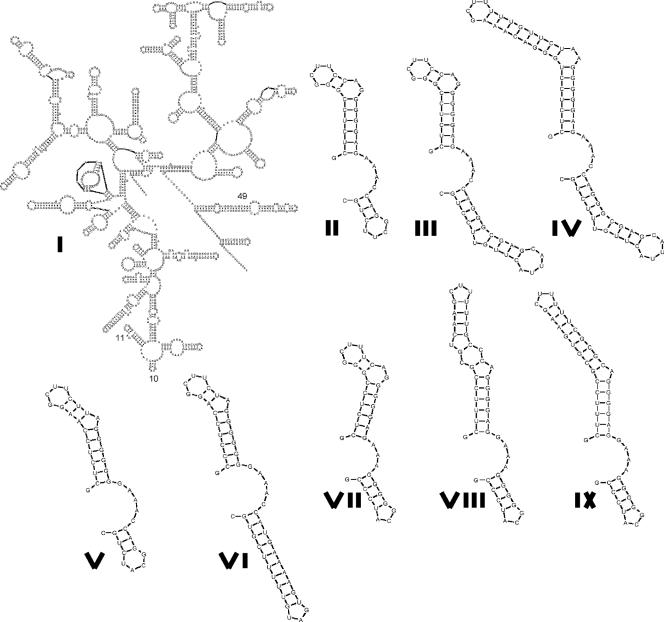
FIG. 3.
Structural comparison of the relevant portions of the 16S rRNAs (helices 10 and 11) of piezophilic and nonpiezophilic strains. Alignments of the same regions are shown in Fig. 2. I, 16S structure of Escherichia coli, with the locations of helices 10, 11, and 49 indicated; II to IV, Photobacterium profundum SS9 helices 10 and 11 from different ribotypes; V, Colwellia psychrerythraea 34H; VI, Colwellia sp. strain MT41; VII, Shewanella oneidensis MR1; VIII, Shewanella benthica PT99; IX, Shewanella benthica KT99.

