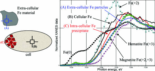
FIG. 3.
XANES analyses of (A) extracellular Fe-rich material (blue), (B) regions of the cell without granules (black), and (C) intracellular granules formed during ferrihydrite reduction (red) (drawing not to scale). The calibration standard for Fe(III) was hematite (Fe2O3) (magenta triangles), that for Fe(II) and Fe(III) was magnetite (Fe3O4) (green squares), and that for Fe(II) was FeCl2 (light-blue inverted triangles).

