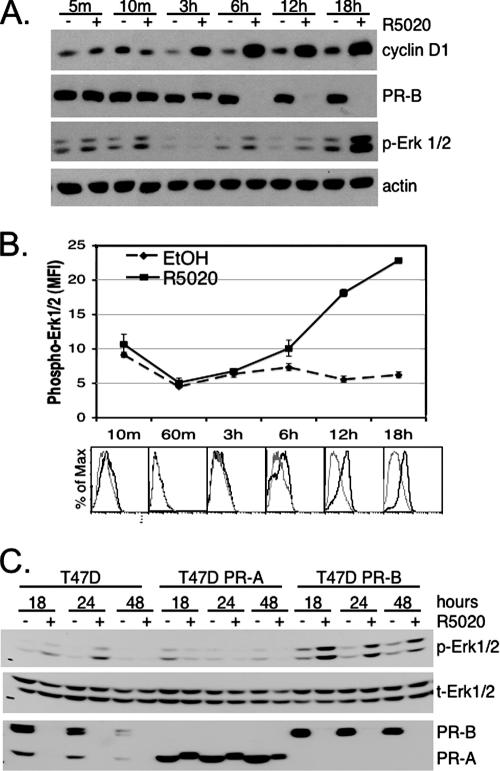
FIG. 1.
Progestin treatment of T47D-YB cells stimulates PR-dependent sustained activation of Erk1/2 MAPKs. (A) T47D cells stably expressing wt PR-B were treated with EtOH vehicle control (−) or 10 nM R5020 (+) for the indicated times (m, minutes). Cell lysates were Western blotted for expression of cyclin D1, PR, or activated (phospho) forms of Erk1/2 MAPKs. Actin blotting was included as a protein-loading control; total MAPK did not change in the same lysates (not shown) (see the legend to panel C). (B) Time course of Erk1/2 phosphorylation as measured using flow cytometry following EtOH control (dashed lines) or R5020 (solid line) stimulation. The lines represent the mean fluorescence intensities (MFI) of triplicate measures versus time; the error bars represent the standard errors of the mean. Histogram overlays (below) at each time point represent fluorescence versus the percentage of maximum for EtOH (gray line) compared to R5020 (black line) treatment. The specificity of the assay was demonstrated using the MEK 1/2 inhibitor U0126 to block R5020-dependent increase in phospho-Erk1/2 (data not shown). The experiments were repeated three times with similar results. (C) T47D parental cells that endogenously expressed both PR-A and PR-B, or sublines engineered to stably express either PR-A or PR-B, were treated with or without R5020 for 18, 24, or 48 h; the cell lysates were Western blotted for phospho- (p) or total (t) Erk1/2 MAPK and total PR. Three independent experiments yielded identical results.

