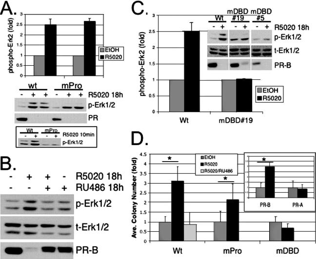
FIG. 5.
Contributions of rapid PR signaling and PR transcriptional events to sustained MAPK activation and anchorage-independent growth. (A) Duplicate cultures of T47D cells stably overexpressing wt PR-B or the c-Src binding mutant, PR-B mPro, were treated with EtOH vehicle control (−) or R5020 (+) for 18 h or 10 min (bottom), and whole-cell lysates were Western blotted for phospho-Erk1/2 (p-Erk1/2) MAPK and total PR. The duplicate 18-h experimental time points shown, from a representative experiment, were quantified by densitometry of visible control and experimental bands on scanned images and expressed as increased Erk2 MAPK activity (n-fold) relative to vehicle controls (the bars represent means plus standard deviations). Densitometry of multiple exposures yielded similar results. Note that Western blotting confirmed the inability of PR-B mPro to rapidly activate Erk1/2 at 10 min relative to wt PR-B (bottom). The results were repeated three times. (B) T47D-YB cells were treated with EtOH, 10 nM R5020, 10 nM R5020 plus 100 nM RU486, or 100 nM RU486 alone for 18 h; the cell lysates were subjected to Western blotting with antibodies specific for phospho- (p) or total (t) Erk1/2 MAPK and total PR. Similar results were obtained for three experiments. (C) Phospho-Erk1/2 MAPK Western blotting and quantification by densitometry of wt-PR-B-expressing T47D-YB cells and two clones (19 and 5) stably expressing the PR-DBD mutant, C587A (mDBD). The cells were treated as described above with vehicle control (−) or R5020 (+) for 18 h, and whole-cell lysates were Western blotted for phospho- or total Erk1/2 and PR-B. PR blotting indicated roughly equivalent levels of PR protein in stable cell lines. The bars represent the R5020-stimulated activation (n-fold; plus standard deviation) of Erk2 MAPK for duplicate measurments performed within the same experiment relative to controls, using stable PR-DBD mutant C587A clone 19 (mDBD). Densitometry of multiple exposures yielded similar results. The experiments were repeated three times with similar results. (D) Progestin-stimulated soft-agar colony formation of T47D cells stably expressing either wt PR-B or PR-B mutants, mPro or mDBD. Cells were plated as described in Materials and Methods, and the bars represent the increase (n-fold) (plus standard deviation) in colony numbers for each stable cell line relative to EtOH controls. The inset shows colony numbers (n-fold) for R5020-treated T47D-YB (PR-B) cells relative to cells stably expressing PR-A (T47D-YA; PR-A). The asterisks denote significance (P < 0.01) determined by an unpaired Student's t test between EtOH- and R5020-treated conditions. The results were confirmed in two independent experiments.

