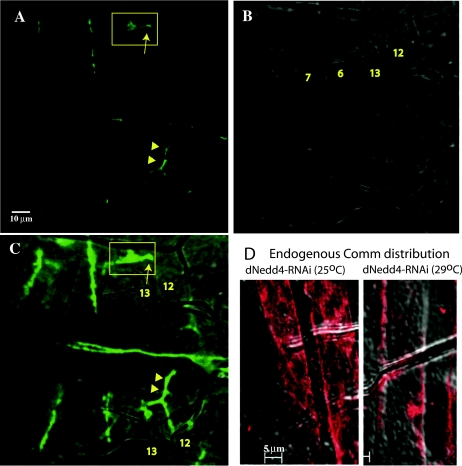
FIG. 8.
Detection of muscle innervation defects along the SNb branch in embryos following knockdown of dNedd4 by RNAi. (A to C) Innervation defects on muscles 12 and 13 in UAS-dNedd4RNAi/24B-GAL4 stage 17 embryos grown at 29°C. Arrow in the boxed areas depict stalling of innervation of muscle 12 compared to normal innervation patterns shown below (arrowheads). (A) Confocal image; (B) DIC image; (C) overlay of the two images. (D) Confocal images (overlaid on a differential interference contrast image) of muscles from stage 17 embryo grown at 25°C (left) or 29°C (right) and immunostained with anti-Comm-ECD. Note the intracellular (intramuscle) distribution of endogenous Comm at 25°C (where there is no knockdown of dNedd4) and surface accumulation of Comm at 29°C (where dNedd4 is knocked down). Scale bars, 10 μm in panel A and 5 μm in panel D.

