FIG. 8.
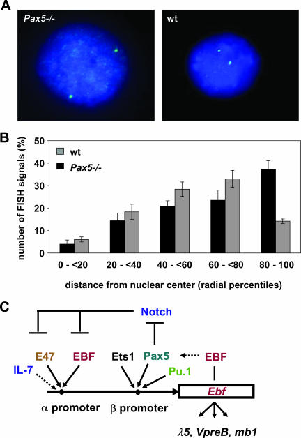
Fluorescence in situ hybridization shows differential Ebf1 locus positioning in Pax5-deficient compared to wild-type (wt) pro-B cells. (A) Representative images of 2D-FISH analysis of Pax5-deficient and wild-type pro-B cells. (B) Analysis of the nuclear position of the Ebf1 locus shows that the Ebf1 locus is more often detected at the periphery of the nucleus in Pax5-deficient than in wild-type pro-B cells. (C) A complex regulatory network regulates the expression of the two Ebf1 promoters.

