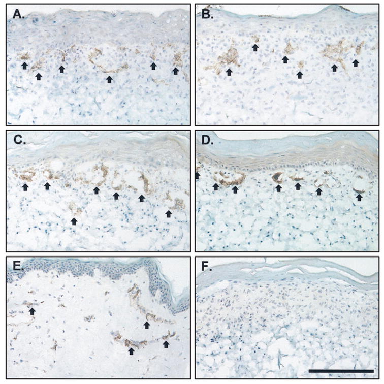
Figure 3.

Staining of human endothelial cells in cultured skin substitutes plus endothelial cells (CSS + ECs) in vitro. Shown is immunostaining for human CD31 (arrows, dark brown staining), an EC marker. A. Control CSS + ECs (group 3), week 1 in vitro. B. VEGF-modified CSS + ECs (group 4), week 1 in vitro. C. Control CSS + ECs (group 3), week 2 in vitro. D. VEGF-modified CSS + ECs (group 4), week 2 in vitro. E. normal human skin. F. CSS without EC (group 2), negative control. The epidermis is at the top of each panel; scale bar in F (0.2 mm) is the same for all panels.
