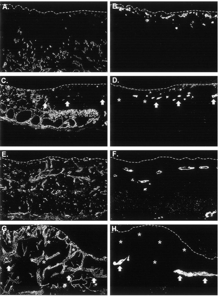
Figure 5.

Proliferation of human endothelial cells after grafting is not enhanced in vascular endothelial growth factor (VEGF)-modified cultured skin substitutes plus endothelial cells (CSS + ECs). Sections were double-labeled with species-specific anti-CD31 antibodies to simultaneously localize mouse (A, C, E, G) and human (B, D, F, H) ECs. A and B. Control CSS + ECs (group 3) 1 week after grafting. C and D. VEGF-modified CSS + ECs (group 4) 1 week after grafting. E and F. Control CSS + ECs (group 3) 3 weeks after grafting. G and H. VEGF-modified CSS + ECs (group 4) 3 weeks after grafting. Dotted white lines indicate the locations of the dermal–epidermal junctions; sections are oriented with the epidermis at the top of each panel. In VEGF-modified CSS + ECs, relatively few human EC clusters (arrows) were observed in regions that were densely populated with mouse vessels (asterisks).
