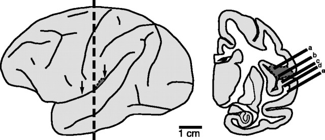
Figure 1.
Recording in the SII region. Lateral (left) and coronal (right) views of the brain are shown. The vertical dashed line indicates the coronal plane of the section. Arrows indicate the approximate AP extent of the SII hand region (dark gray area), which is located mainly in the UBLS. Right, Possible microelectrode penetrations made in and around the SII region. Penetrations a and b encountered neurons that were driven by somatosensory and sometimes visual stimulation, whereas penetrations c-e encountered neurons that were driven by auditory stimulation.

