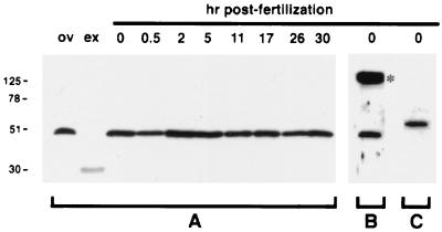
Figure 5.
Western blot analysis of concentrated egg exudate and whole-egg lysates separated under reducing conditions. Positions of molecular mass markers (in kilodaltons) are on the left. (A) A blot probed with anti-ovochymase antibodies, with lanes representing ovary lysate (ov), concentrated exudate of activated dejellied eggs (ex), and lysates of intact fertilized eggs collected at the given times after fertilization. (B) A blot of unactivated egg lysate probed with anti-ovotryptase1 antibodies; the asterisk marks an interference band representing large amounts of egg yolk protein (28). (C) A blot of unactivated egg lysate probed with anti-ovotryptase2 antibodies.

