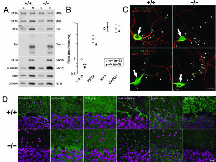
Fig. 4.
Altered distribution of KIF1 in ROSA22 mutant neurons and mice. (A) Equal amounts of soma (S) and neurite (N) lysates prepared from wild-type (+/+) and ROSA22 mutant (−/−) mice were subjected to Western blot analyses, with antibodies against proteins indicated. (B) Signal intensities of bands of KIF1A, 3A, 5, and GAPDH were quantified, and the data are presented as the ratio of the amount in neurites to that in soma. The values presented are mean ± SEM of three independent experiments. ∗∗, P < 0.01 with paired t test. (C) In wild-type (+/+) neurons, EGFP-KIF1A is localized in the soma (white arrow) and entered neurites (small white arrowheads). In neurons from ROSA22 homozygotes (−/−), EGFP-KIF1A was present in the soma (white arrow) but was absent from neurites (white arrowheads). In contrast, EYFP-KIF5A (white arrowheads) was able to enter neurites in neurons from both wild-type and ROSA22 homozygous mice. Green, kinesins; red, MAP2. (Scale bar, 20 μm.) (D) Sagittal brain sections of control (+/+) and ROSA22 mutant (−/−) mice were stained with antibodies that recognize the proteins indicated (green) and TOTO-3 (magenta). Note that the distribution of Tau and MAP1A appears to be altered in the ROSA22 mutant cerebellum. (Scale bar, 20 μm.)

