Fig. 3.

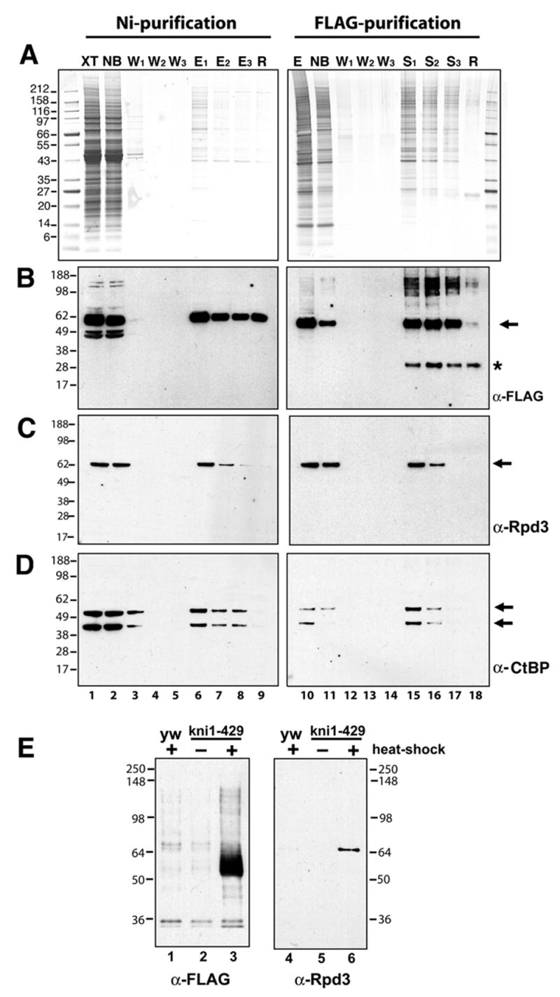
Double affinity purification of Knirps protein reveals copurification of CtBP and Rpd3 proteins. Knirps protein was purified by sequential binding to Ni-NTA affinity resin and anti-FLAG antibody coupled to protein G sepharose beads. A, Coomassie (left) and silver (right) stained gels showing protein composition of crude lysate (XT, lane 1), nonbound material (NB, lane 2), wash fractions 1–3 (W, lanes 3–5; lanes 1–5, 0.007% of total), imidazole eluates from Ni-NTA beads (E1–3, lanes 6–8, 0.13% of total) and material retained on beads after elution (R, lane 9). Panel on the right shows the pooled Ni-NTA eluates (E, lane 10), material that did not bind to anti-FLAG antibody sepharose (NB, lane 11), three washes (W, lanes 12–14; lanes 10–14, 0.16% of total), material eluted from antibodies with Sarkosyl (S, lanes 15–17), and material remaining on beads (R, lane 18; lanes 15–18, 0.08% of total). B, Western blot with anti-FLAG antibody reveals presence of Knirps protein (arrow) during fractionation. Only a fraction of Knirps in the crude lysate is retained on Ni-NTA, however, most of the Ni-NTA purified Knirps subsequently binds to the anti-FLAG beads. Asterisk indicates nonspecific band. C, Western blot with anti-Rpd3 antibodies. A portion of this protein cofractionates with Knirps through the two affinity purification steps. D, Western blot with anti-CtBP antibody reveals fractionation of this corepressor; both short and long forms of the protein copurify with recombinant Knirps protein. E, E, After double affinity purification, Rpd3 is present exclusively in fractions containing recombinant Knirps. Shown are Western blots of proteins from heat-shocked non-transgenic embryos, or from uninduced or heatshock induced transgenic embryos. Double affinity purifications were performed in parallel using equivalent amounts of embryos. Left panel: no anti-FLAG staining material (Knirps protein) is found in fractions from control embryos (lanes 1–2), while a strong band is found in fractions from extracts containing induced Knirps protein (lane 3). Right panel: Rpd3 protein is recovered only in fractions from induced, Knirps expressing embryos (lane 3) and not from controls (lanes 1–2).
