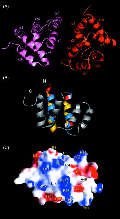
Figure 3.
Models of the Apaf-1/caspase-9 CARD complex. (A) Model of Apaf-1 CARD/caspase-9 CARD binary complex. Apaf-1 CARD is colored in pink, whereas caspase-9 CARD is colored in brown. The structure of caspase-9 CARD is constructed based on homology modeling of Apaf-1 CARD by using segment matching method (33). (B) Ribbon representation of caspase-9 CARD. The acidic, basic, and hydrophobic residues of α1 and α4 are colored in red, blue, and yellow, respectively. (C) Surface diagram of caspase-9 CARD in the same orientation as in B. In this figure, the surface electrostatic potential is color coded such that regions with electrostatic potentials <−8 kBT are red, whereas those >+8 kBT are blue (where kB and T are the Boltzmann constant and temperature, respectively). Surface-exposed hydrophobic residues are labeled in black.

