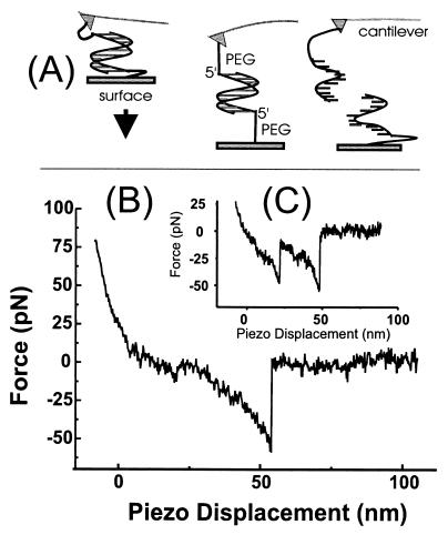
Figure 1.
Measurement of unbinding forces. (A) The complementary single strands of a DNA are immobilized on an AFM tip and a surface via their 5′-ends by PEG linker molecules. On approach of the surface to the tip, a duplex may form that is loaded on retract until an unbinding occurs. (B) A typical force-versus-piezo displacement for the DNA duplex a⋅b during the retract of the sample with a velocity of 100 nm/s shows that, after initial contact of the tip with the surface (positive force values), the DNA duplex is loaded and the 30 nm long PEG-linker molecules are stretched (negative force values). At a displacement of ≈50 nm, the duplex unbinds at a loading of 50 pN. (C) A force displacement curve in which two molecules unbind one after the other, the last unbinding event also being at ≈50 pN loading.

