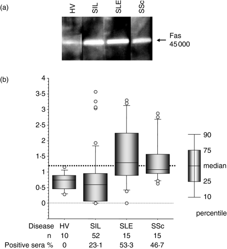
Figure 1.
Detection of anti-Fas autoantibodies by Western blot analysis. (a) Human Fas linked to glutathione-s-transferase was separated by 10% sodium dodecyl sulphate–polyacrylamide gel electrophoresis and electrotransferred to a polyvinylidene difluoride membrane. The blots were incubated with sera (1/100 dilution) from either a healthy volunteer (HV) or patients with silicosis (SIL), systemic lupus erythematosus (SLE) or systemic sclerosis (SSc). The blot was then incubated with horseradish peroxidase-conjugated sheep anti-human immunoglobulin (1/15 000 dilution). The bound antibodies were detected using the enhanced chemiluminescence method. (b) The ratio of the intensity of the detected band in the patient samples was calculated relative to that of a healthy volunteer (HV) analysed at the same time. The cut-off point was determined to be 1·2 by the receiver operating characteristic curve (indicated by the thick dotted line). The percentage of positive sera for each patient is also shown. Open circles represent outlier values.

