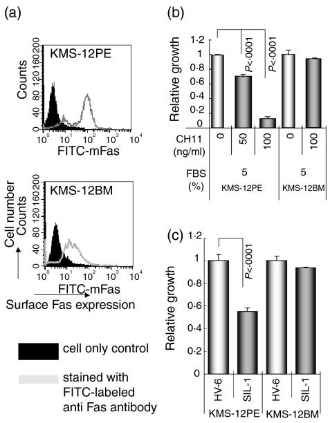
Figure 5.
(a) Surface Fas expression levels of KMS-12PE and KMS-12BM human myeloma sister cell lines. The filled histogram indicates the cell only control and the open histogram indicates cells stained with fluorescein isothiocyanate-labelled anti-Fas monocloal antibody. (b) The relative growth of KMS-12PE and KMS-12BM cells cultured with RPMI-1640 medium plus 5% FBS with or without (control) the Fas-mediated apoptosis inducing CH11 antibody (50 and 100 ng/ml) as analysed by the WST-1 assay with the control value being set at 1·0. Although growth of the Fas-expressing KMS-12PE cell line was inhibited by CH11, growth of KMS-12BM cells showed no change. (c) KMS-12PE and KMS-12BM cell lines were cultured with serum from HV-6 or SIL-1 (the SIL-1 serum contained a large amount of anti-Fas autoantibodies as analysed by Western blotting and ProteinChip System). The serum from SIL-1 inhibited growth of KMS-12PE, a Fas-expresser, but not KMS-12BM, a low Fas-expresser.

