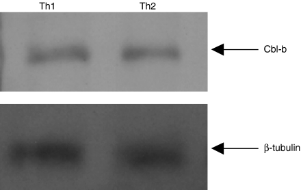
Figure 2.
Equivalent Cbl-b protein expression levels in Th1 and Th2 cells. Th1 and Th2 (2 × 10⁶) cells were lysed in ice-cold buffer containing 25 mm Tris, pH 7·5, 150 mm NaCl, 5 mm ethylenediaminetetra-acetic acid, 1 mm phenylmethylsulphonyl fluoride, 1 mm Na3VO4, 1 µg/ml aprotinin, 1 µg/ml leupeptin and 1% NP-40. The proteins were separated on a 10% sodium dodecyl sulphate–polyacrylamide gel electrophoresis and immunoblotted with anti-Cbl-b antibody and developed using ECL Western blotting detection system (Amersham, UK). The blot was stripped and reprobed with anti-β-tubulin antibody. The data are representative of two individual experiments.

