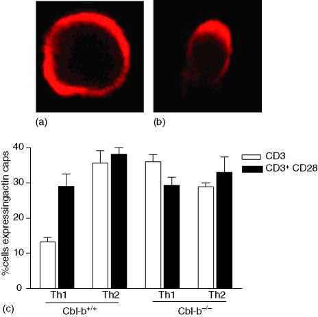
Figure 4.
Cbl-b deficiency induces actin reorganization in Th1 cells. Th1 and Th2 cells derived from Cbl- b +/+ and Cbl-b–/– mice were stimulated with CD3 or CD3/CD28 antibody coated polysytrene beads. Actin polymerization was visualized by phalloidin staining using confocal microscopy. Cell:bead interactions were counted. Cells displaying even staining all around were considered negative for actin capping (a) and actin polymerization was defined as capping or aggregation of phalloidin staining at one pole of the cell (b). The percentage of cells with actin caps was calculated as (actin capping/total cell:bead interactions) × 100 (c) Data are mean ± SD from three separate experiments.

