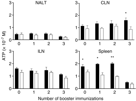
Figure 5.
Responses of cells isolated from the nasal-associated lymphoid tissue (NALT), cervical lymph nodes (CLNs), iliac lymph nodes (ILNs) and spleen following one, two or three booster immunizations. Animals not boosted (zero boost) were used as a control group. Animals were killed 8 days after final booster immunization or at the 7-month time-point for those not boosted. Cells were cultured with surface antigen AgI/II (solid bars) or culture medium alone (open bars), and proliferation was determined by quantifying cellular ATP concentrations from individual cultures. The significance of differences was evaluated between AgI/II-stimulated and unstimulated cultures. *P < 0·05; **P < 0·01.

