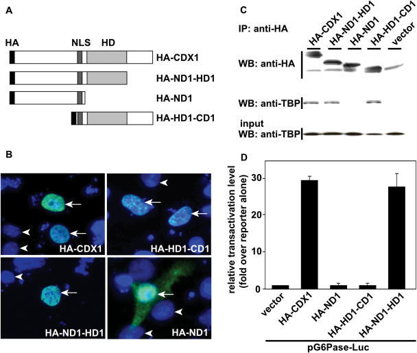
Figure 3.
Requirement of different domains of CDX1 for TBP-binding and transactivation. (A) Schematic representation of the truncated forms of CDX1. HA: hemagglutinin epitope, NLS: nuclear localization signal, HD: homeodomain. (B) Intracellular distribution of the recombinant proteins. HCT116 cells were transfected with the plasmids encoding the indicated proteins and immunolabeled with primary rabbit anti-HA antibody followed by secondary anti-rabbit immunoglobulins coupled to Alexa 488. Cell nuclei were stained with DAPI. Arrows denote cells expressing the mutant forms of CDX1; arrowheads indicate nuclei of non-transfected cells labeled only with DAPI. (C) Co-immunoprecipitation of TBP with the truncated forms of CDX1. HCT116 cells were co-transfected with the plasmids encoding TBP and either HA-CDX1, HA-ND1-HD1, HA-ND1, HA-HD1-CD1 or the control empty vector. Proteins immunoprecipitated with anti-HA antibody were detected by western blots using anti-HA and co-immunoprecipitation of TBP was assayed using anti-TBP. TBP was recovered in every fraction except with HA-ND1. The presence of TBP in the cell extracts prior to HA-immunoprecipitation was controlled by western blot using anti-TBP antibody (Input). (D) Transcriptional activity of the truncated forms of CDX1. HepG2 cells were co-transfected with the reporter plasmid pG6Pase-Luc and with each of the expression vectors encoding HA-CDX1, HA-ND1-HD1, HA-ND1 or HA-HD1-CD1, together with the control reporter pRL-null. The data obtained in triplicate ±SD are representative of three independent experiments.

