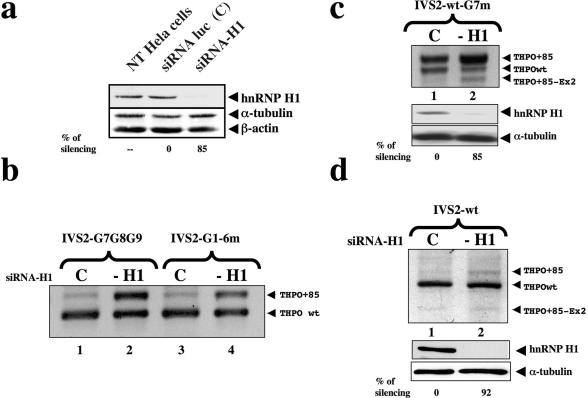
Figure 6.
Knock-down of hnRNPH1 causes the activation of the cryptic acceptor site within IVS2. (a) Western blot carried out with polyclonal anti-hnRNP H1 antiserum on HeLa cell non transfected (NT HeLa cells) or transfected with siRNA against hnRNP H1 (siRNA-H1) or luciferase (siRNA-luc). β-tubulin and α-actin were probed with polyclonal antibodies as a control of total protein loading. Relative amounts of hnRNP H1 silencing are indicated below each lane. Standard deviations were always ≤20%. (b) The panel shows splicing pattern analysis of RT–PCR products after cotransfection of IVS2 G7G8G9 and IVS2-G1-6m along with either the siRNA-H1 (lanes 2 and 4) or a control siRNA-luc oligonucleotides (lanes 1 and 3). (c) RT–PCR experiments after RNAi against hnRNP H1 and cotransfection with the IVS2-wt-G7m minigene. Lane 1 shows the cotransfection of IVS-wt-G7m with a control siRNA. After siRNA anti-hnRNP H the THPO+85 isoform became predominant (lane 2). Lower panels show the related anti-hnRNP H1 western blot analyses. β-tubulin was used as a control of total protein loading. Relative amount of hnRNP H1 silencing is indicated below each lane. Standard deviation were ≤20%. (d) RT–PCR experiments in siRNA-H1-treated in HeLa cells. Lanes 1 and 2 show the results of PCR after cotransfecting the IVS2-wt minigene. After the reduction in the expression of hnRNP H there is a small increase of the THPO+85 isoform. The control transfection was loaded in lane 1. Lower panels show the related anti-hnRNP H1 western blot analyses with indication of relative amounts of hnRNP H1 silencing. Standard deviations were ≤20%.

