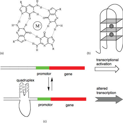
Figure 1.
(a) Structure of a G-tetrad, showing hydrogen bonds and monovalent cation. (b) Schematic of an intramolecular G-quadruplex. G-tetrads are shown as blue squares, and monovalent cations as grey spheres. The structure shown is folded in an antiparallel conformation, with the strands of the G-tetrads alternately running up and down. (c) Model for transcription modulation via formation of a quadruplex in a promoter region.

