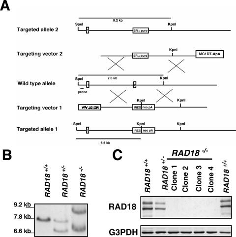
Figure 1.
Generation of RAD18−/− HCT116 cells. (A) Schematic representation of RAD18 locus, disruption constructs and configuration of targeted alleles. Relevant restriction sites and the position of the probe used for Southern blot analysis are shown. MC1, MC1 promoter; DT-ApA, diphtheria toxin gene; puro, puromycin resistance gene; neo, neomycin resistance gene; SRα, SRα promoter; IRES, internal ribosome entry site; pA, poly (A) additional signal. Exons 1 and 2 are indicated by numbered boxes. (B) Southern blot analysis of KpnI- and SpeI-digested genomic DNA from cells with indicated genotypes of RAD18 gene. (C) Western blot analysis of whole-cell extracts from different HCT116 clones with indicated genotypes of RAD18 gene. G3PDH was used as an internal control.

